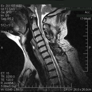
Cervical Myelopathy
The cervical spondylotic myelopathy is defined as a compression of the cervical spinal cord due to degenerative changes in the cervical spine.
Cervical myelopathy surgery
Both anterior and posterior approaches had been used as a surgical treatment for cervical myelopathy.
Anterior Cervical Decompression can be performed, depending on the severity of the case, through multiple discectomies and performing ACDF (Anterior Cervical Discectomy (Decompression) & Fusion) and in selected cases, a corpectomy can be performed at one or more levels with patient bone graft (ilium or fibula).
For all surgical techniques, it requires a rigorous patient selection and precise canal decompression to enforce a neurological improvement. It is important to mention that perioperative complications can be devastating in this type group of high risk patients with cervical spondylotic myelopathy, but with careful attention, meticulous technique and surgical experience, excellent results can be achieved.
Intraoperative Monitoring (IOM) of the Nerves
We usually recommend to perform Intraoperative Monitoring (IOM) of the Nerves in Cervical Myelopathy Surgery.
Frequency of segments that can be affected
C4-C5: 2%
C5-C6: 19%
C6-C7: 69%
C7-D1: 10%
Myelopathy diagnosis
X-rays (Radiology): it can show signs of cervical spondylosis, but gives no confirmation of myelopathy by itself.
CT (computed tomography): It is more accurate when focusing on the amount of cord compression, as it is far better than an MRI when evaluating the bone.
MRI (Magnetic Resonance Imaging): It is the most selected method for patients with cervical myelopathy. Besides giving an assessment of the degree of spinal canal stenosis, MRI can identify myelomalacia or permanent spinal cord lesions.
How is cervical Myelopathy produced?
Due to the aging of the vertebral discs and later on, height loss and dry out of the discs, causes a reduction in diameter of the spinal canal and spinal cord compression. And subsequently osteophytes take place, that is, calcifications stick out from the edges of the surfaces of the vertebral bodies, which produce both dorsal and ventral compression of the spinal cord. Symptoms develop when the spinal canal is reduced at least by 30%.
Moreover, the normal movement of the cervical spine can aggravate spinal cord damage due to mechanically direct compression. During flexion, the spinal cord is stretched, and it is being stretched on the ventral osteophytes. During the extension, the yellow ligament already calcified causes a reduction of space available for the cord to flex. Over time, a lack of blood flow causes spinal cord ischemia and thus myelopathy takes place.
Cervical myelopathy symptoms
The symptoms of this disorder are characterized by a slow decline, stepwise, with signs worsening in gait disturbance, weakness, sensory disturbances and usually some degree of pain. Diagnosis can often be made based on the findings of the patients’ medical history, physical examination and with plain radiographs, but is it necessary to undergo an MRI for a complete confirmation.
Slight symptoms without clear manifestations of gait disorders or pathological reflexes require no surgical treatment, but patients with demonstrable myelopathy and cord compression are most likely candidates for surgery.
The main clinical symptoms of myelopathy and cervical spondylotic radiculopathy include primarily by the following:
– Painful neck or arms
– Sensitive disorders
– Motor disorders (including the incapacity of fine movements)
Clinically, myelopathy cases predominantly show lower limb weakness, slurring of fine movements (the coordination of small muscle movements) and gait disturbance. Contrariwise, the presence of radicular pain without legs involvement suggests a cervical radiculopathy, which is a dysfunction of a nerve root of the cervical spine also known as pinched nerve.
Sources:
Dr. Vicenç Gilete, MD, Neurosurgeon & Spine Surgeon.
Neurosurgery volumes I–III. Edited by Robert H. Wilkins and Setti S. Rengachary. McGraw-Hill.
Handbook of Neurosurgery. Mark S.Greenberg, Seventh Edition. Thieme.
Last Update [site_last_modified date_format=”Y-m-d H:i:s”]